CCL Injuries
Understanding the Anatomy of Your Pet's Knee
If your dog has been diagnosed with a torn ACL (anterior cruciate ligament) then most likely your veterinarian has suggested some form of surgical correction for this problem. Your veterinarian has probably told you that you have surgical options and may have referred you to an orthopedic specialist to have surgery performed.
Unfortunately, we veterinarians sometimes speak in ways that leave you confused, making it difficult for you to make appropriate decisions for your pet. I would like to try to explain to you, in plain English, what is involved in an ACL (anterior cruciate ligament) injury and its repair.
We need define our terms so we can understand the problem better.
In canine patients, the ACL is properly called a CCL, or cranial cruciate ligament. The term ACL refers to the same structure, but in humans. Your veterinarian may refer to it as an ACL because most people have heard of athletes with torn ACLs and understand what part of the anatomy we are speaking of. From here on out, I'll be using the proper term, cranial cruciate ligament, CCL.
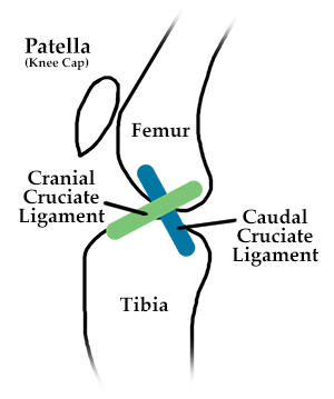
 The knee joint (the joint where the CCL resides) is properly called the stifle joint. The stifle joint is composed of three major bones: the femur, the tibia, and the patella. In humans, those three are your thighbone (femur), shin bone (tibia) and knee cap (patella). The ends of these bones are surrounded by cartilage, the slippery stuff that allows for movement. They sit together in a fluid called joint fluid, and there is a seal around the joint called a joint capsule.
Inside the joint sits the CCL and another ligament called the caudal cruciate ligament. They form a cross shape, hence the name cruciate (meaning cross). This is important later when explaining joint function.
Also in the joint are two shock absorbers called meniscus. When you see an X-ray of your pet's stifle joint, the femur and tibia appear to be separated by space, but they are not. The femur sits on top of the meniscus. The meniscus is not visible on an X-ray. For that matter, ligaments are not visible on an X-ray either, so you cannot see the CCL on an X-ray.
Let's now explain the function of the stifle joint:
The stifle joint is a complex joint; but I will simplify it so that its function can be understood. First, compare the stifle to a hinge that can only swing two ways, forward and backward. The center of the hinge is inside of the stifle joint. The forward swing is called extension, and occurs during weight bearing. The backward swing is called flexion and occurs during the non-weight bearing, or swing, phase of gait motion.
As everyone knows, when you walk, only one foot is on the ground at any one time. Now most importantly, the cranial and caudal cruciate ligaments keep the femur (thighbone) in alignment with the tibia (shin) during motion. These two ligaments, in the shape of an X, or cross, maintain appropriate contact between the two bones.
The CCL attaches in the back of the femur and comes forward to attach to the front of the tibia. The caudal cruciate ligament attaches on the front of the femur, and goes backwards to attach to the back of the tibia. Where they cross each other is the "hinge" point.
Understanding the Injury: CCL Rupture
 What happens when the CCL ruptures?
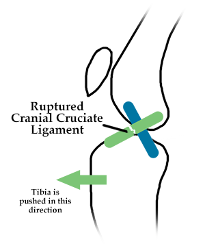
We've all had a door break a hinge, and then the door won't open or close very well, usually causing a lot of scraping on the floor. The same thing happens in the stifle joint. The bones are no longer in proper alignment, and the joint doesn't work well, causing pain, inflammation and cartilage damage.
Typically, when the CCL ruptures, the femur rides backwards on the tibia, and the tibia wants to come forward. The fancy term for this phenomenon is cranial tibial subluxation. The bottom line is that it is very painful for your pet.
Most clients bringing in a pet with a CCL rupture say that it occurred during running, fetching or playing with another dog. To the owner, it may appear as an acute (or quick onset) injury, but that is not so. Unlike humans, this is a chronic disease in dogs. Human ACL injuries are almost always the result of athletic trauma.
You may ask, "What's the difference between human and canine knee ligament injuries?"
First, the stance of the canine knee is different than the stance of the human knee. People stand straight up, with their femur directly on top of their tibia, having a joint angle of 1800. Dogs stand with an angle of 1350. Every time that a dog stands, the bone alignment is dependent upon an intact CCL to hold the bones in place in the stifle (knee) joint. Chronic wear and tear on the CCL causes it to ultimately fail.
Your pet's CCL is like a rope made up of many fibers. With time and stress, the fibers of the rope slowly break down until it is so weak that it can't do its job anymore and breaks.
You can also have a partial tear which is just as painful, and leaves the CCL just as incompetent. This is important because CCL injury is always accompanied by some form of degenerative joint disease or osteoarthritis. This arthritis is not reversible.
Most surgeries slow the progression of arthritis, but do not reverse what is already there; therefore the stifle is never the same as if it had an intact CCL. (The world is not perfect!)
Understanding Your Surgical Options
The surgeries for your pet's torn CCL fall into two broad categories:
- Procedures that attempt replacement of the broken CCL
- Procedures that change the geometry of the stifle so that the CCL is not needed.
Replacing the Broken CCL
The surgeries that replace the broken ligament are further divided into two major categories:
- intracapsular repair
- extracapsular repair
The capsule refers to the joint capsule, or seal, that surrounds the stifle joint.
The intra-capsular repair uses the body's natural material to recreate the CCL. This procedure is commonly done in human medicine, but has fallen to the wayside in veterinary medicine, as the results have not been as good as in other procedures. I won't perform an intracapsular repair, but stay tuned, some researchers are looking at synthetic material inside the joint, and this surgery may make a comeback.
The extracapsular repair is a common form of CCL repair. There are many different ways and materials available to stabilize the stifle joint. This is where much confusion occurs, as all of these procedures have different names, but the theory is the same: re-create the CCL outside of the joint.
Of these procedures, the only one to use the body's natural material is a procedure called the lateral fibular head transposition, which is a fancy name for re-creating the CCL with the lateral collateral ligament. All of the other forms of extracapsular repair use synthetic materials such as polyester, nylon, or proprietary materials (meaning that it is a patented material).
All of these procedures have two things in common:
- they attach on the side of the femur, and
- come forward to attach to the front side of the tibia.
Terms you may hear your veterinarian use to describe the procedures include among others:
- lateral suture stability
- modified Flo
- femoral tibial suture
- Tight Rope™
- Bone Biters™
Some veterinarians drill tunnels, some use anchors, and some pass around another bone. They are all on the outside of the stifle joint. These procedures are commonly done on smaller dogs, and can be quite successful. They do have their share of problems, however, especially with larger dogs.
Big dogs can break their real CCL ligament, and they can certainly break or stretch the fake one. As this artificial material is placed on the outside surface of the joint, normal joint mechanics are inhibited.
During the stance or weight bearing phase, the tibia bone rotates a small amount in a normal dog, but this internal rotation is inhibited by the artificial material placed outside of the joint. The procedures that we talked about above can still can lead to a non-lame dog, and may be recommended for smaller dogs or older non-active dogs; however, their shortcomings have led to newer procedures that modify the stifle joint geometry.
Making the Joint Work Without the CCL
Geometry is the study of angles and forces, and these newer procedures use that study to modify joint function by changing angles inside the stifle joint, so that the CCL is not necessary.
These procedures differ greatly from those that attempt to re-create the CCL. The two most common procedures are the TPLO (tibial plateau leveling osteotomy), and the TTA (tibial tuberosity advancement). Both accomplish the same thing: cutting the tibia bone, making a bony shift and using a bone plate to hold the cut piece of bone in place.
Tibial Plateau Leveling Osteotomy (TPLO)
The TPLO, first developed in the mid-1990s, is explained by dissecting the name. The tibia is the bone involved: tibial = bottom bone of the stifle joint. Plateau is the name for the top of the tibia bone inside the joint, where the femur sits. Leveling is in relation to the floor, giving the femur a level spot to sit on the tibial plateau. Osteotomy is a fancy term meaning to cut bone. (The actual angles measured and cut are not to the floor of the surgery room, but this simplifies a basic understanding.)
Tibial Tuberosity Advancement (TTA)
Tibial tuberosity advancement (TTA) is also explained by its name. Tibial = relating to the tibia, the lower half of the of the stifle joint. The tuberosity is the front edge of the tibia just below the stifle joint where the patellar tendon attaches, and advancement, meaning to bring forward. The tibial tubersosity (patellar tendon attachment) is moved forward and a spacer is screwed in place to hold it together until the bone cut is healed.
When the tibial tuberosity is moved to a 90o angle, the stifle joint is stabilized by shifting the weight to the caudal cruciate ligament. In effect there is no longer a need for the cranial cruciate ligament.
If you wish to read in further detail on the mechanics of the TTA procedure, visit the inventor's website: www.kyonpharma.com. There you will find an exhaustive description of the forces in play on stifle mechanics and how the TTA works. The diagrams are very well done but can be confusing due to the complexity of the stifle joint.
What is the best procedure for my dog's CCL rupture?
That question can only be answered on an individual basis, as each patient is unique and many factors including anatomy and lifestyle come into play when making a decision. Although both TTA and TPLO preocedures are highly successful, some surgeons will have a preference for one or the other in certain situations.